Watch the exceptional presentations, plus the Q&A panel discussions, on demand from Symposium 2023!

Maximize the Detection of Pathogenic Aberrations With Optical Genome Mapping

Hematological malignancies are characterized by multiple classes of chromosomal aberrations. Comprehensive detection of these aberrations is vital to properly classify samples and provide actionable information. Due to the limitations of current cytogenetic approaches, only a fraction of the samples evaluated lead to a positive aberration finding11-13. Optical genome mapping (OGM) significantly improves detection to unlock truly comprehensive chromosomal aberration profiles and maximize the number of informed cases.

Achieve, with a single assay, detection of aberrations from all classical cytogenetic technologies combined.

  • With a single workflow, find all classes of chromosomal aberrations detected by karyotyping, FISH, and chromosomal microarrays combined
  • Generate results with >99% concordance with standard cytogenomic methods as demonstrated by multiple peer-reviewed studies
  • Achieve 10,000x resolution and higher sensitivity compared to karyotyping, with a scalable digital approach


Find more pathogenic and actionable chromosomal abnormalities.

  • Reveal significantly more pathogenic aberrations than classical cytogenetics, with a higher-resolution, genome-wide, and unbiased method
  • Enable more pathogenic discoveries, resolve cryptic cases, and determine complex events like chromothripsis with much higher precision and resolution
  • Increase the number of informed cases, and reduce the rate of cases signed off as “normal” due to lack of findings from classical methods

Simplify and transform your lab operations.

  • Transform your lab operations by reducing complexity and number of assays required per sample
  • Implement a simpler sample-to-answer workflow that does not require cell culture and has more automated steps with simpler data analysis
  • Leverage sophisticated software solutions to visualize and call variants, annotate and interpret findings, and report results

Curated Publications

See the research global experts are publishing on OGM and hematological malignancies.
Read More

“The combination of OGM and a targeted NGS panel for genome profiling of myeloid cancer is more cost effective than 60x whole-genome sequencing and provides the most comprehensive genome analysis.”

Dr. Ravindra Kolhe
Augusta University

“30% of previously unsolved cases for B-ALL, which previously underwent karyotype + FISH + microarray + NGS, were solved using OGM.”

Dr. Gordana Raca
Children’s Hospital Los Angeles

Data Examples

Multiple peer-reviewed studies from global experts have demonstrated the same outcomes: OGM has a high correlation to results from traditional methods across diverse types of hematological malignancies, while consistently revealing additional pathogenic findings.

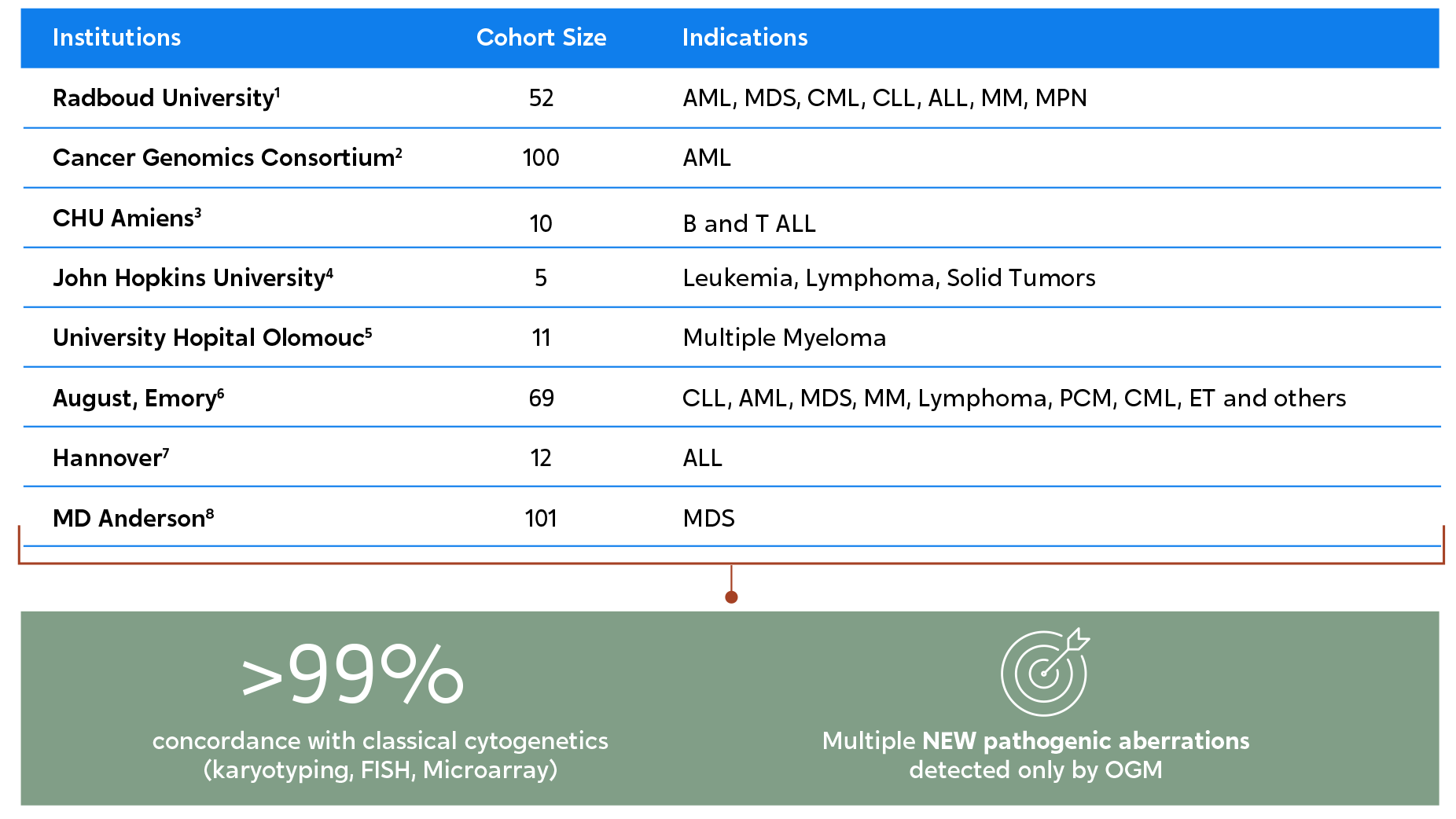
When compared to traditional cytogenetic methods, OGM can uncover up to twice as many pathogenic chromosomal aberrations.

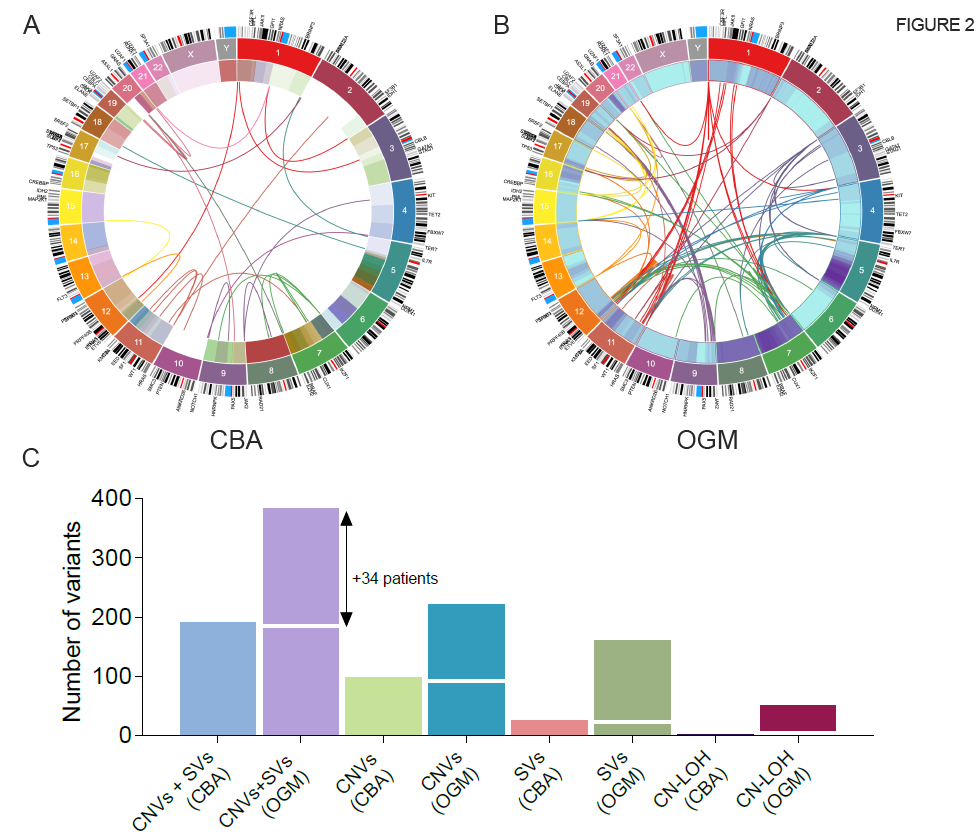
In a 2022 MD Anderson Cancer Center study, OGM detected twice the number of clinically significant chromosomal aberrations as karyotyping in 101 myelodysplastic samples.8 This figure illustrates the Circos plot view of the variants identified by karyotyping (left) versus OGM (right).

High-resolution and enhanced characterization of samples by OGM also leads to better risk analysis and stratification of samples.


Disease Impact of OGM Analysis
AML and MDS9 For 67% of cases, the karyotype was redefined. In some cases, this led to an adjustment in ELN risk classifications
AML and MDS10 11% of samples for which OGM findings led to change in R-IPSS or 2010/2017 ELN score
MDS8 17% of cases assessed with OGM had the IPSS-R risk reclassified from initial karyotyping

Discover How OGM Benefits Clinical Research in Cancer

Sign up to watch the AMP 2022 Bionano Corporate Workshop #1, which discusses Maximizing Detection of Pathogenic Structural Variants Across Hematological Malignancies with Optical Genome Mapping.
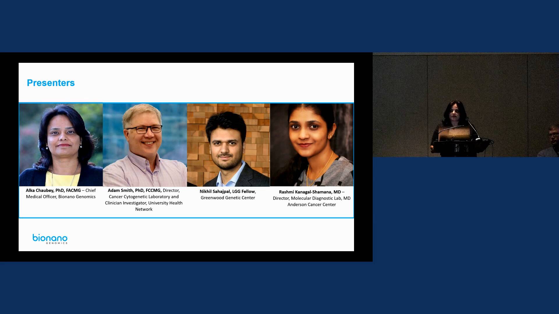
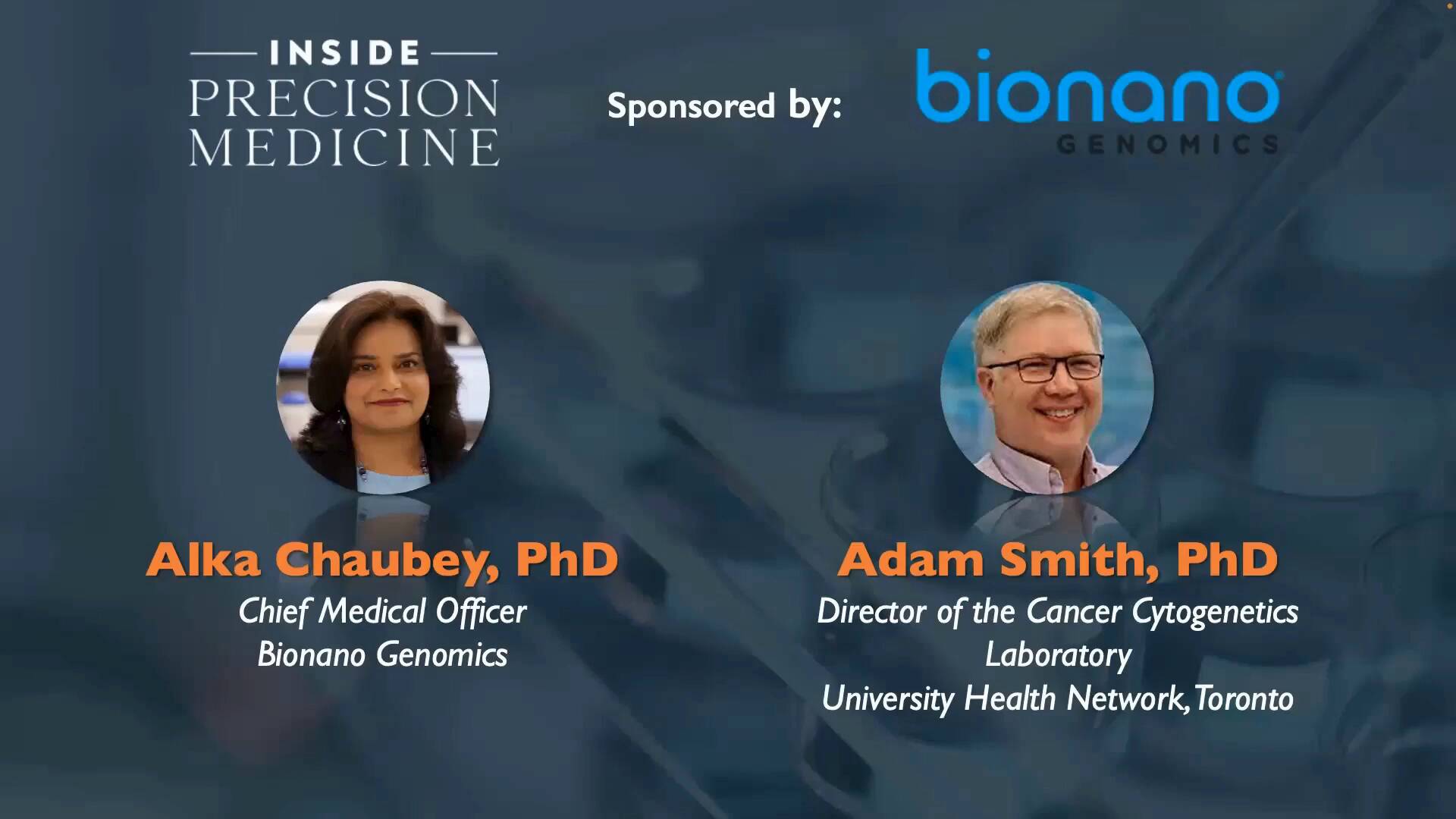
Watch Videos

Sign up to watch this webinar and hear from Bionano’s Dr. Alka Chaubey and Dr. Adam Smith, UHN, as they discuss how Optical Genome Mapping Meets Challenges for New Blood Cancer Classifications
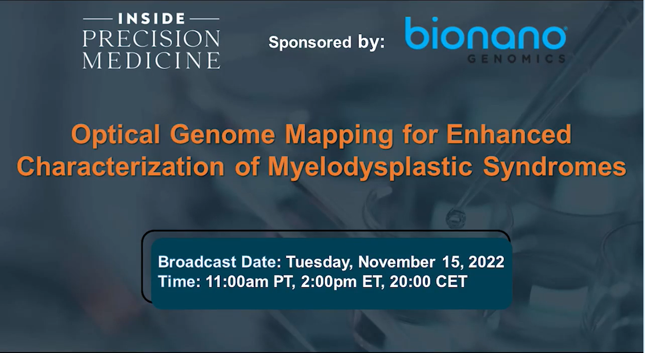
Watch On-demand

Sign up to watch this webinar and hear how OGM makes a huge impact on clinical research, as Dr. Rashmi Kanagal-Shamanna and Dr. Guillermo Garcia-Manero (MD Anderson Cancer Center) discuss OGM for Enhanced Characterization of Myelodysplastic Syndromes.

Watch On-demand

Learn More About OGM

Read about what structural variations are and why they matter.

Learn More

See how OGM reveals structural variation in a way that has never been done before.

Learn More

Find the latest research in our Publications Library.

Learn More

Sign up to get the latest updates on Bionano solutions for heme applications.

Sign Me Up!

RELATED MATERIALS

title Source Authors
Optical Genome Mapping Identifies Novel Recurrent Structural Alterations in Childhood ETV6::RUNX1+ and High Hyperdiploid Acute Lymphoblastic Leukemia September 21, 2023

Brandes, Danielle; Yasin, Layal; Nebral, Karin; Ebler, Jana; Schinnerl, Dagmar; Picard, Daniel; Bergmann, Anke; Alam, Jubayer; Köhrer, Stefan; Haas, Oskar A.; Attarbaschi, Andishe; Marschall, Tobias; Stanulla, Martin; Borkhardt, Arndt; Brozou, Triantafyllia; Fischer, Ute; Wagener, Rabea

Assembly of 43 diverse human Y chromosomes reveals extensive complexity and variation June 12, 2023

Pille Hallast, Peter Ebert, Mark Loftus, Feyza Yilmaz, Peter A. Audano, Glennis A. Logsdon, Marc Jan Bonder, Weichen Zhou, Wolfram Hoeps, Kwondo Kim, Chong Li, Philip C Dishuck, David Porubsky, Fotios Tsetsos, Jee Young Kwon, Qihui Zhu, Katherine M. Munson, Patrick Hasenfeld, William T. Harvey, Alexandra P. Lewis, Jennifer Kordosky, Kendra Hoekzema, The Human Genome Structural Variation Consortium (HGSVC), Jan O. Korbel, Chris Tyler-Smith, Evan E. Eichler, Xinghua Shi, Christine R Beck, Tobias Marschall, Miriam K. Konkel, Charles Lee

Babesia duncani multi-omics identifies virulence factors and drug targets April 13, 2023

Pallavi Singh, Stefano Lonardi, Qihua Liang, Pratap Vydyam, Eleonora Khabirova, Tiffany Fang, Shalev Gihaz, Jose Thekkiniath, Muhammad Munshi, Steven Abel, Loic Ciampossin, Gayani Batugedara, Mohit Gupta, Xueqing Maggie Lu, Todd Lenz, Sakshar Chakravarty, Emmanuel Cornillot, Yangyang Hu, Wenxiu Ma, Luis Miguel Gonzalez, Sergio Sánchez, Karel Estrada, Alejandro Sánchez-Flores, Estrella Montero, Omar S. Harb, Karine G. Le Roch, Choukri Ben Mamoun

  1. Neveling K, Mantere T, Vermeulen S, et al. Next-generation cytogenetics: Comprehensive assessment of 52 hematological malignancy genomes by optical genome mapping. The American Journal of Human Genetics. 2021;108(8):1423-1435. doi:10.1016/j.ajhg.2021.06.001
  2. Levy B, Baughn LB, Chartrand S, et al. A national multicenter evaluation of the clinical utility of optical genome mapping for assessment of genomic aberrations in Acute myeloid leukemia. December 2020. doi:10.1101/2020.11.07.20227728
  3. Lestringant V, Duployez N, Penther D, et al. Optical genome mapping, a promising alternative to gold standard cytogenetic approaches in a series of acute lymphoblastic leukemias. Genes, Chromosomes and Cancer. 2021;60(10):657-667. doi:10.1002/gcc.22971
  4. Stinnett V, Jiang L, Haley L, et al. Adoption of optical genome mapping in Clinical Cancer Cytogenetic Laboratory: A stepwise approach. Cancer Genetics. 2022;260-261:3. doi:10.1016/j.cancergen.2021.05.021
  5. Kriegova E, Fillerova R, Minarik J, et al. Whole-genome optical mapping of bone-marrow myeloma cells reveals association of extramedullary multiple myeloma with chromosome 1 abnormalities. Scientific Reports. 2021;11(1). doi:10.1038/s41598-021-93835-z
  6. Sahajpal NS, Mondal AK, Tvrdik T, et al. Clinical validation and diagnostic utility of optical genome mapping for enhanced cytogenomic analysis of hematological neoplasms. The Journal of Molecular Diagnostics. November 2022. doi:10.1016/j.jmoldx.2022.09.009
  7. Lühmann JL, Stelter M, Wolter M, et al. The clinical utility of optical genome mapping for the assessment of Genomic Aberrations in acute lymphoblastic leukemia. Cancers. 2021;13(17):4388. doi:10.3390/cancers13174388
  8. Yang H, Garcia-Manero G, Sasaki K, et al. High-resolution structural variant profiling of myelodysplastic syndromes by optical genome mapping uncovers cryptic aberrations of prognostic and therapeutic significance. Leukemia. 2022;36(9):2306-2316. doi:10.1038/s41375-022-01652-8
  9. Gerding W, Tembrink M, Nilius-Eliliwi V, et al. Optical genome mapping reveals additional prognostic information compared to conventional cytogenetics in AML/MDS patients. Int J Cancer. 2022 Jun 15;150(12):1998-2011. doi: 10.1002/ijc.33942.
  10. Balducci E, Kaltenbach S, Villarese P, et al. Optical genome mapping refines cytogenetic diagnostics, prognostic stratification and provides new molecular insights in adult MDS/AML patients. Blood Cancer J. 2022;12(126). doi: https://doi.org/10.1038/s41408-022-00718-1
  11. Tsui SP, Ip HW, Saw NY, et al. Redefining prognostication of de novo cytogenetically normal acute myeloid leukemia in young adults. Blood Cancer J. 2020 Oct 19;10(10):104. doi: 10.1038/s41408-020-00373-4. PMID: 33077814; PMCID: PMC7573626.2. Nimer. Best Pract Res Clin Haematol. 2008. PMID: 18342811.
  12. Nimer SD. Is it important to decipher the heterogeneity of “normal karyotype AML”? Best Pract Res Clin Haematol. 2008 Mar;21(1):43-52. doi: 10.1016/j.beha.2007.11.010. PMID: 18342811; PMCID: PMC2654590.
  13. Walker A, Marcucci G. Molecular prognostic factors in cytogenetically normal acute myeloid leukemia. Expert Rev Hematol. 2012 Oct;5(5):547-58. doi: 10.1586/ehm.12.45. PMID: 23146058; PMCID: PMC3582378.4. Graessner et al. Eur J Hum Genet. 2021. PMID: 34140650.